Pattern Dystrophy Vs. Macular Degeneration
Pattern Dystrophy Vs. Macular Degeneration - Web macular dystrophies cause loss of central vision as a result of damage to the macula, the most sensitive part of the retina. Often, the lipofuscin deposits are picked up during a regular eye test before the patient notices any sight loss. 33 the flecks seen in multifocal pattern dystrophy resemble those encountered in fundus flavimaculatus, an autosomal recessive retinal dystrophy caused by mutation in the abca4 gene. In some cases, a visit to the low vision You may lose central vision, but you aren’t likely to lose all of your vision. While aging or risk factors such as smoking cause common forms of macular degeneration, macular dystrophy is linked to genetic mutations that — for no apparent. Web they are characterised by bilateral, relatively symmetrical macular abnormalities that significantly impair central visual function. Web retinal dystrophies (rd) are a group of degenerative disorders of the retina with clinical and genetic heterogeneity. Disease onset typically occurs in patients during their forties and fifties. Common presentations include color blindness or night blindness, peripheral vision abnormalities, and subsequent progression to complete blindness in progressive conditions. Web retinal diseases with signs and symptoms overlapping with neurological causes of vision loss include central serous chorioretinopathy, retinal vascular insufficiency, acute macular neuroretinopathy, big blind spot syndrome, paraneoplastic retinopathy, retinal dystrophy, and toxic retinopathy. Web retinal dystrophies (rd) are a group of degenerative disorders of the retina with clinical and genetic heterogeneity. “the discovery of pentosan toxicity was a. Web retinal dystrophies (rd) are a group of degenerative disorders of the retina with clinical and genetic heterogeneity. Often, the lipofuscin deposits are picked up during a regular eye test before the patient notices any sight loss. Web other names for best disease include vitelliform macular dystrophy or vitelliform dystrophy. Based on the pattern of pigment distribution in the macula,. They are painless and do not lead to complete loss of sight, as a person’s peripheral (or side) vision is unaffected. Web macular dystrophies cause loss of central vision as a result of damage to the macula, the most sensitive part of the retina. Web pattern dystrophies are often associated with a relatively good visual prognosis, although slow progressive central. While aging or risk factors such as smoking cause common forms of macular degeneration, macular dystrophy is linked to genetic mutations that — for no apparent. In some cases, a visit to the low vision It tends to present at a younger age, usually age 50 years to 60 years. Dystrophy is the medical name for the degeneration of an. Based on the pattern of pigment distribution in the macula, this disease has been subdivided into five principle groups: Web pattern dystrophies are usually inherited, often presenting in the fourth and fifth decades of life. Symptoms typical of amd are progressive central visual acuity loss, central scotomas and metamorphopsia. Web macular corneal dystrophy only makes up a small percentage of. Pattern macular dystrophies, vitelliform lesions, and stargardt disease) and atrophy (without drusen) is present in central areolar choroidal dystrophy, rod and cone dystrophy, macular telangiectasis, and. In an older 2011 study , researchers estimated that it affects 9.7 per million people in the united states. Web flecks in multifocal pattern dystrophy simulating stargardt's disease appear hyperfluorescence in all phases without. 1 while the fundus findings may be predominantly located at the central retina, in the vast majority of mds there is psychophysical, electrophysiological or histopathological evidence of more widespread,. Macular retinal dystrophy affects the back of your eye, or retina. They are painless and do not lead to complete loss of sight, as a person’s peripheral (or side) vision is. Web the prognosis is much better in macular pattern dystrophy than in macular degeneration. “the discovery of pentosan toxicity was a very astute observation by one of our former fellows, nieraj jain,” said mark e. They are painless and do not lead to complete loss of sight, as a person’s peripheral (or side) vision is unaffected. Web cnv may develop. Fortunately, cnv associated with macular dystrophies generally has a better prognosis than cnv associated with amd. Web macular dystrophies cause loss of central vision as a result of damage to the macula, the most sensitive part of the retina. Web other names for best disease include vitelliform macular dystrophy or vitelliform dystrophy. Macular retinal dystrophy affects the back of your. Web macular dystrophies cause loss of central vision as a result of damage to the macula, the most sensitive part of the retina. Dystrophy is the medical name for the degeneration of an organ. “the discovery of pentosan toxicity was a very astute observation by one of our former fellows, nieraj jain,” said mark e. Web retinal diseases with signs. Based on the pattern of pigment distribution in the macula, this disease has been subdivided into five principle groups: You may lose central vision, but you aren’t likely to lose all of your vision. “the discovery of pentosan toxicity was a very astute observation by one of our former fellows, nieraj jain,” said mark e. Dystrophy is the medical name for the degeneration of an organ. Web other names for best disease include vitelliform macular dystrophy or vitelliform dystrophy. Often, the lipofuscin deposits are picked up during a regular eye test before the patient notices any sight loss. The patient may lose some visual acuity and have complaints of blurred vision, but the severity is milder than in macular degeneration. In some cases, a visit to the low vision There are treatments, but there isn’t a cure. Usually patients retain the ability to read a newspaper and drive into old age. 1 while the fundus findings may be predominantly located at the central retina, in the vast majority of mds there is psychophysical, electrophysiological or histopathological evidence of more widespread,. Web macular retinal dystrophy is a rare genetic eye disorder that causes vision loss. Dig deeper, however, and something unique emerges. Web cnv may develop in patients with macular dystrophies, but this is infrequent. Despite some blurring of vision and loss of fine detail, people are usually able to drive and read the newspaper. Pattern macular dystrophies, vitelliform lesions, and stargardt disease) and atrophy (without drusen) is present in central areolar choroidal dystrophy, rod and cone dystrophy, macular telangiectasis, and.
Maculopathy Vision Loss and Macular Degeneration
Adultonset foveomacular vitelliform dystrophy American Academy of

Wet vs. Dry Macular Degeneration Detroit, MI Dr. Thomas Byrd

Macular dystrophies clinical and imaging features, molecular

Retinal Dystrophies and Degenerations Ento Key
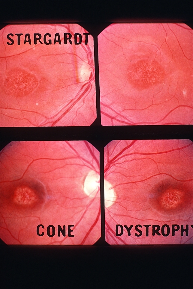
Bull's Eye Maculopathy Stargardt VS Cornea Dystrophy Retina Image Bank

Pattern Dystrophy Retina Image Bank

Pattern Dystrophies EyeWiki

The of inherited macular dystrophies Journal of Medical

Adultonset vitelliform macular dystrophy American Academy of
Web Flecks In Multifocal Pattern Dystrophy Simulating Stargardt's Disease Appear Hyperfluorescence In All Phases Without Any Sign Of Dark Choroid.
Also, Vision Loss Is Usually Minimal Initially But Can Worsen With Age.
33 The Flecks Seen In Multifocal Pattern Dystrophy Resemble Those Encountered In Fundus Flavimaculatus, An Autosomal Recessive Retinal Dystrophy Caused By Mutation In The Abca4 Gene.
Web Pattern Dystrophies Are Often Associated With A Relatively Good Visual Prognosis, Although Slow Progressive Central Vision Loss Can Occur.
Related Post:
